Our equipment
Our values decide what we buy. When a problem is hard to solve — a cornea that will not correct, a child whose short-sightedness is moving faster than it should, an eye surface that has stopped being comfortable — the answer usually depends on measuring something most practices cannot measure. So we invest in the instruments that can, and we keep investing as the technology moves.
We have top of the line equipment to accurately diagnose and monitor eye conditions, treat specific eye conditions and fit specialty contact lenses. It means we can provide continuity of care without referring elsewhere for testing — and it is why patients travel to us from across New Zealand and overseas, and why other practitioners refer to us for co-management.

Why we invest in this
Our values decide what we buy. When a problem is hard to solve — a cornea that will not correct, a child whose short-sightedness is moving faster than it should, an eye surface that has stopped being comfortable — the answer usually depends on measuring something that most practices cannot measure. So we invest in the instruments that can, and we keep investing as the technology moves.
Rose Optometry has been in Frankton since 1969. Paul Rose's specialty was keratoconus, and what came out of that work was the Rose K lens — designed here in Hamilton, launched in 1990, and now the most frequently prescribed lens in the world for the irregular cornea, manufactured in 19 countries and distributed in over 95. Paul was made a Companion of the New Zealand Order of Merit for it: the first ophthalmic professional in New Zealand to receive that honour. Keratoconus care did not arrive at this practice. It started here.
That is the practice people come to. Patients travel to us from all over New Zealand, and most weeks of the month there is at least one from overseas — among them patients who fly in from Ethiopia, Europe, Dubai, China, Japan and the United States. Not for the brands on our equipment list, but for the fact that a difficult eye gets measured properly here, by people who look at these problems every week. The instruments exist to serve that. They are the reason we can say yes to cases other practices have to decline.
For other eye care practitioners
It is also why other eye care practitioners send patients to us. We accept referrals every week from optometrists across the Waikato and further afield — Specsavers, OPSM, Bailey Nelson, Visique, For Eyes and independent practices alike — for co-management of keratoconus, complex contact lens fitting, myopia control, dry eye and anterior eye disease. A referral is not a transfer. The patient goes back to their own optometrist with a plan, and we stay available for the parts that need our instruments. If you are a practitioner reading this, please contact us for co-management; we are here to help.
Research sits separately
Research is held in the New Zealand Eye Research Centre rather than in the practice. That separation is deliberate. Clinical trials have their own protocols, their own timelines and their own paperwork, and none of that should ever slow down or reshape an ordinary appointment at Rose. Keeping the two apart is how the practice stays a premium clinical service while the research centre does research.
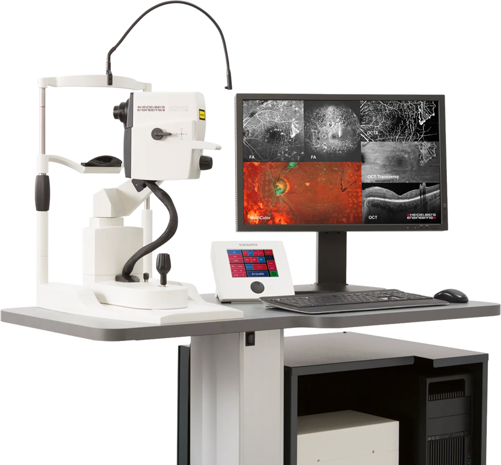
The OCT (Optical Coherence Tomographer) machine of choice for ophthalmologists and optometrists alike. Used to image a number of structures in the eye, the Spectralis OCT sees deep into the layers of the retina and optic nerve in high resolution, imaging layers that cannot be detected with other OCT machines – much like an MRI. This allows us to pick up some eye conditions earlier than when using other machines, and monitor them more effectively using the highly detailed images and the progression tools within the software. The machine can also be used for some contact lens fitting, helping our optometrists to achieve the best fit for your eyes.
Ours is not a standard instrument. Three additions do things a base OCT cannot.
Nsite Analytics. A neuro-ophthalmic module that measures the retinal nerve fibre layer and the ganglion cell layer as a marker of neurological change, not only ocular change. Heidelberg describe it as providing a window into the brain, because those layers are central nervous system tissue that can be photographed directly. It is the analysis used in multiple sclerosis and neurodegenerative disease research. To the best of our knowledge, no other optometry practice in New Zealand has this module.
BluePeak blue-laser autofluorescence. Rather than injecting a dye, this images the eye’s own natural fluorescence, which comes from the retinal pigment epithelium. Cells that are stressed light up; cells already lost go dark. It maps disease at a cellular level in macular degeneration and inherited retinal disease, non-invasively, in seconds — and it routinely shows the true extent of a problem well beyond what is visible on a retinal photograph. Scanning laser angiography sits on the same platform for the cases where dye imaging is genuinely required.
Heidelberg AppWay, and NMI-ChoroidAI. AppWay is Heidelberg’s application marketplace, which means the instrument gains new analysis without new hardware. The application that matters most to us is NMI-ChoroidAI, developed by NetraMind Innovations. It takes an ordinary Spectralis volume scan and automatically segments the choroid in three dimensions, reporting choroidal thickness, choroidal volume and the choroidal vascularity index. The choroid thins as an eye elongates and thickens when a myopia treatment is working, so it is an early signal of response — and it moves before a prescription does.
What this means for you: an OCT scan is painless, takes seconds and touches nothing. Its real value is comparison — this year’s scan measured against last year’s, which is how glaucoma, macular degeneration and diabetic change are caught while they are still small. Scanning is included where indicated in a New Patient examination (45 minutes) or an Existing Patient examination (30 minutes). If we have asked you to repeat imaging, book an OCT (30 minutes) or Fields and OCT together (45 minutes).
For colleagues: if you are managing myopic children with an OCT but no biometer, please contact us for co-management — choroidal measurement can give you an objective response marker from the instrument already in your practice.
The gold standard of optical biometry. Rose Optometry is the only optometry practice in Waikato with an IOL master; a high-precision measurement tool used to measure the axial length of the eye, corneal curvature, and anterior chamber depth with exact detail. The IOL master allows us to monitor progression of myopia (short sightedness) with impeccable accuracy and assess the efficacy of myopia control treatment. It is an integral part of our myopia control clinic.
What this means for you: axial length is a far better measure of a child's myopia than the glasses prescription alone, because it tracks the eye's actual growth. If your child is short-sighted, start with a free Myopia Chat (15 minutes, no cost), or read more on our myopia control page. Existing patients due for a repeat biometry measurement can book an IOL Master appointment (15 minutes).
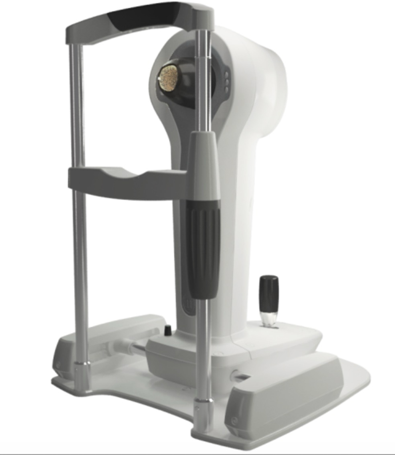
The topographer of choice used by most specialists, universities and researchers. Rose holds six units — a Medmont in every consulting room — so corneal mapping happens during your appointment rather than being deferred to a separate visit. The newer Meridia units add high-resolution imaging of the eye surface and the meibomian glands to the curvature map.
A topographer measures the shape of the front surface of the cornea across thousands of points and turns it into a map. Curvature that is steepening in one area, an irregular surface, or an asymmetry between the two eyes are all visible on a map long before they are obvious in a prescription.
What this means for you: corneal topography is how we diagnose and monitor keratoconus, and how we design Ortho-K and specialty lenses to your eye rather than to an average. It is performed inside those appointments, so there is no separate scan to book. New keratoconus patients — book a 45-minute assessment. Curious about Ortho-K — book a free 15-minute chat.
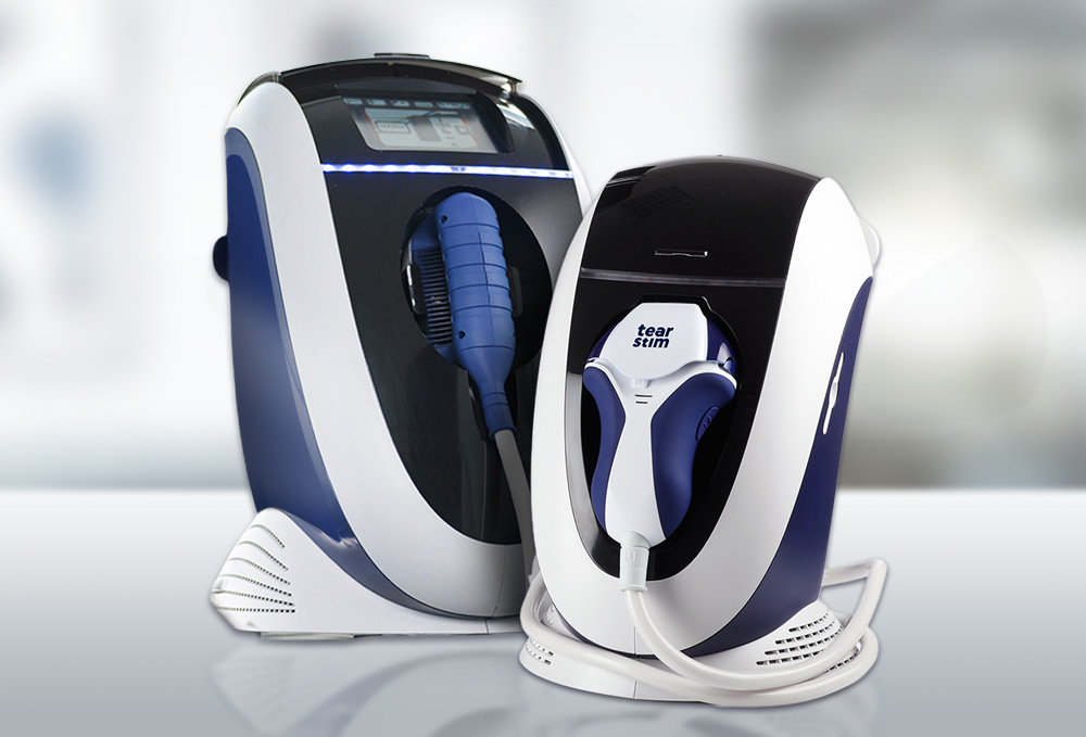
The medical solution to treat dry eye. The French-made E-Eye IPL machine is a clinically proven treatment for dry eye caused by Meibomian Gland Dysfunction, resulting in up to 3 years of symptom relief. 85% of patients report satisfaction after treatment. At Rose Optometry we use the E-Eye in conjunction with Blephasteam goggles to deliver efficient and effective dry eye management at the practice.
What this means for you: IPL treats the cause of evaporative dry eye rather than masking the symptoms, but it is not right for every eye and every skin type. Suitability is decided at an assessment first. Start with a Dry Eye Evaluation, or if IPL has already been recommended to you, book a New IPL assessment (30 minutes). Read more about IPL for dry eye.
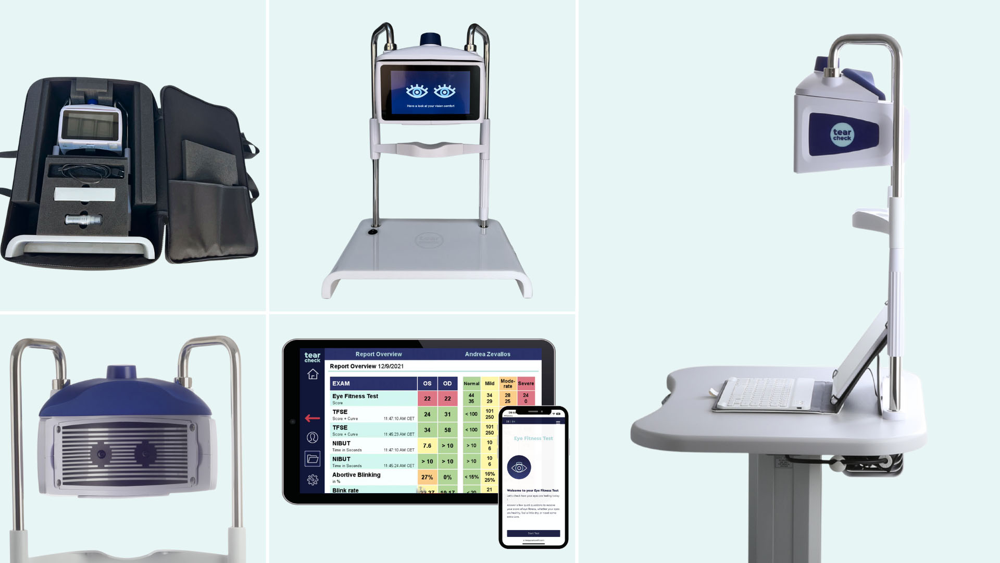
A revolutionary tool for measuring and assessing 9 factors of dry eye. Rose Optometry is the only optometry practice to have the French-made tearcheck available for use on patients. It allows our dry eye specialist optometrists to offer the best tailor made treatments for dry eye without referring elsewhere.
What this means for you: we do not choose a dry eye treatment until we have measured your tear film and your meibomian glands, because gritty, burning or watery eyes have several different causes and the treatments are not interchangeable. TearCheck is used in our Dry Eye Evaluation — a paid, in-depth assessment, with the fee confirmed when you book. Read more about our dry eye clinic.
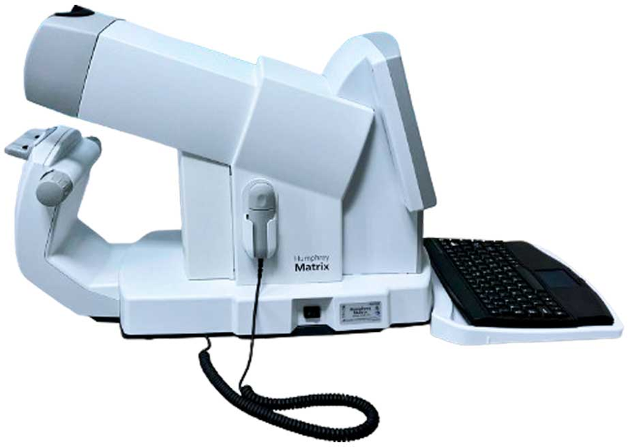
Measures the sensitivity of the visual field (peripheral vision) using Frequency Doubling Technology to pick up visual field defects faster than standard perimetry can. Essential for the diagnosis and management of glaucoma but also useful to assess vision changes due to stroke, tumours, and other neurological conditions.
What this means for you: field loss is usually silent — the brain fills in the gap, so people rarely notice until a great deal has gone. That is why we measure rather than ask. A field test is included where indicated in a full examination: New Patient (45 minutes) or Existing Patient (30 minutes). If we have asked you to repeat a field, book a Visual Field Assessment (15 minutes) or Fields and OCT together (45 minutes).
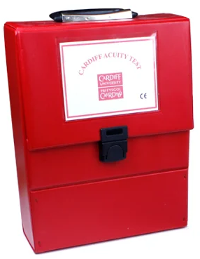
Developed at Cardiff University, Cardiff Acuity Cards measure how well a baby or toddler actually sees — without a single letter, number or spoken answer. Each card carries a simple picture that fades as it gets smaller; the child looks towards the picture while it is still visible to them, and stops looking once it is not. The optometrist watches the eyes, not the mouth. It is the standard way to put a real acuity figure on an infant's vision, and it works from a few months of age. We also hold preferential-looking and picture-matching tests for the ages in between, so a two-year-old, a four-year-old and a seven-year-old are each measured with something suited to them.
What this means for you: Cardiff Cards are not general-purpose optometry equipment. A practice buys them, and learns to use them, because it has chosen to do paediatrics properly — which makes “do you have Cardiff Cards?” a fair question to ask anyone before you take a baby for an eye test. We examine children from six weeks of age. Read more on our children's eye care page, or book a New Child/Student appointment (30 minutes).
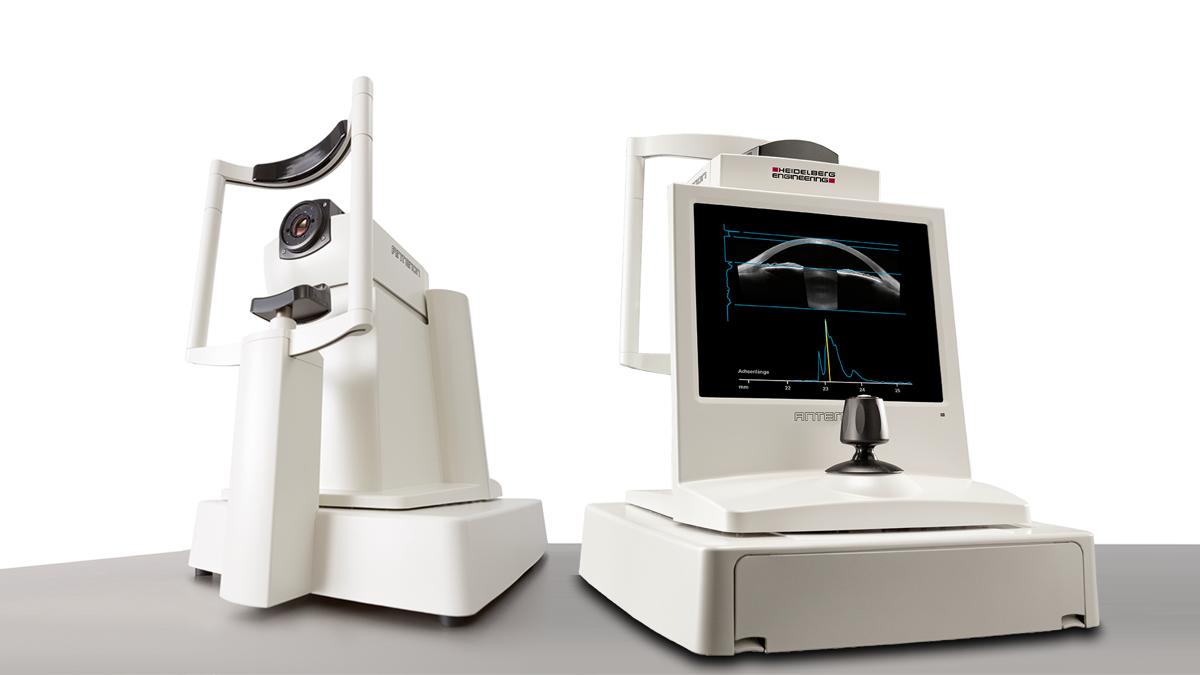
Swept-source OCT of the front of the eye. In one sitting the ANTERION produces corneal tomography (the shape of both the front and the back surface), full-thickness pachymetry across the whole cornea rather than at a single point, epithelial thickness mapping, and biometry for lens and contact lens planning.
Epithelial thickness mapping is the part clinicians care about most. The epithelium is a living surface that remodels to smooth out irregularity beneath it, so in early keratoconus it thins over the developing cone and thickens in a ring around it. That change can appear before the surface shape has altered enough for a topographer to flag it — which is why epithelial mapping can catch keratoconus at the stage where cross-linking protects the most tissue.
The ANTERION also reports the SCORE — Screening Corneal Objective Risk of Ectasia — which combines six measurements into a single figure describing how far a cornea sits from normal. We use it as one input among several, alongside comparison against your own baseline. We also say openly that the published validation of the SCORE cut-offs was carried out in a Korean population, and New Zealand’s keratoconus population is not that population. Closing that evidence gap for New Zealand eyes is exactly the sort of question the New Zealand Eye Research Centre exists to answer.
What this means for you: keratoconus is now diagnosed by numbers rather than by impression, and the numbers change before your vision does. What matters clinically is not any single figure but the direction of travel — a stable cornea and a progressing one can look identical on the day. Cross-linking protects what is still there; it does not restore what has gone, which is the whole argument for scanning early and scanning again. New keratoconus patients — book a 45-minute assessment. Read more about keratoconus.
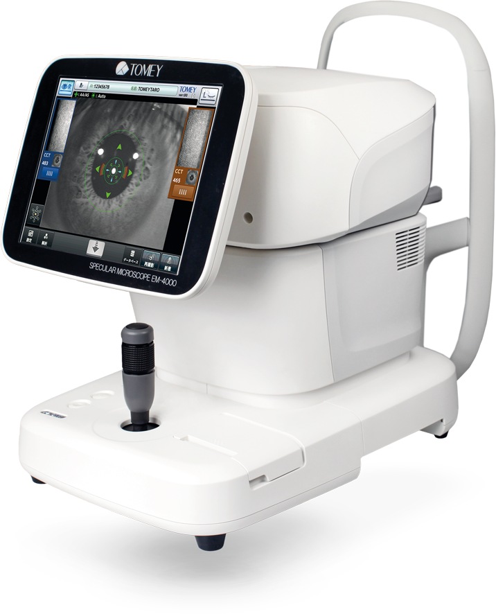
Photographs and counts the endothelial cells on the back surface of the cornea — the single layer of cells that pumps fluid out of the cornea and keeps it clear. You are born with all of them you will ever have and they do not regenerate, so the count only goes one way.
What this means for you: this measurement matters if you wear contact lenses long term, if you have had eye surgery or are considering it, if you have Fuchs’ dystrophy or a corneal graft, or if you are being assessed before cataract surgery. It is quick, touches nothing, and gives a number that can be tracked over the years. It is performed inside a New Patient examination (45 minutes) or a specialty contact lens appointment where indicated.
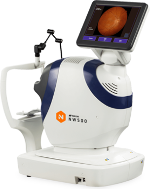
A non-mydriatic retinal camera: it photographs the back of your eye in high resolution without the drops that blur your vision for the rest of the day. The photograph records the optic nerve, the macula and the retinal blood vessels exactly as they were on the day.
What this means for you: a retinal photograph is a permanent record, and its value grows with time. Diabetic change, macular change and optic nerve change are all judged by comparison against a previous image, so the picture taken today is the reference for every year after it. Retinal imaging is included where indicated in a New Patient examination (45 minutes) or an Existing Patient examination (30 minutes).
The reference-standard instrument for measuring the visual field — how much you can see out to the sides, above and below, while looking straight ahead. It is the test used in the clinical trials that define how glaucoma is managed, and its progression software compares each test against your own previous results rather than against an average.
What this means for you: visual field loss is silent. The brain fills in the missing area so convincingly that people rarely notice until a great deal has gone, which is why we measure it rather than ask about it. A field test is included where indicated in a New Patient examination (45 minutes) or an Existing Patient examination (30 minutes). To repeat a field, book a Visual Field Assessment (15 minutes) or Fields and OCT together (45 minutes).
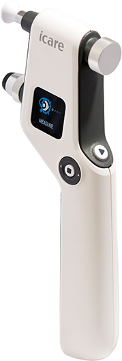
Measures the pressure inside the eye using a probe so light that no anaesthetic drop is needed and most people do not feel it. There is no air puff.
What this means for you: eye pressure is the main modifiable risk factor in glaucoma, so it has to be measured — but the way it is measured decides whether a patient will sit still for it. This instrument is the reason we can take a reliable pressure on children, on anxious patients, and on people who have never managed the air puff test. Pressure is checked as part of a New Patient examination (45 minutes) or an Existing Patient examination (30 minutes).
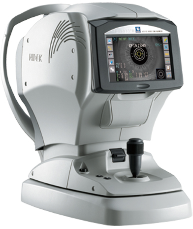
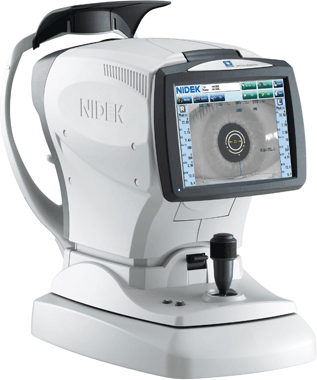
A combined instrument that measures the focusing power of the eye, the curvature of the cornea and the eye pressure in one sitting. It gives the optometrist an objective starting point for your prescription before a single question is asked about which lens looks clearer.
What this means for you: the objective measurement is why an eye test does not depend entirely on your answers. It is particularly valuable for children, for anyone who finds the “which is better, one or two” questions difficult, and for anyone whose prescription has been unstable. These measurements are taken as part of a New Patient examination (45 minutes) or an Existing Patient examination (30 minutes).
A second optical biometer, sitting alongside our ZEISS IOL Master. It measures the axial length of the eye — how long the eye is from front to back — along with corneal curvature, anterior chamber depth and pupil size.
Holding two independent biometers matters more than it sounds. Myopia management turns on hundredths of a millimetre, and a decision to change a child’s treatment should not rest on a single reading from a single machine. Where a measurement looks unexpected, we can cross-check it on a second instrument before acting on it.
What this means for you: axial length is a far better measure of a child’s short-sightedness than the glasses prescription alone, because it tracks the eye’s actual growth against normal growth curves. That is how we see a child drifting off their expected curve before the prescription changes. If your child is short-sighted, start with a free Myopia Chat (15 minutes, no cost), or read more on our myopia control page.
For colleagues: most practices in New Zealand manage myopic children without a biometer, which makes it very hard to know whether a treatment is working or a child simply had a quiet year. If that is you, please contact us for co-management — we are glad to measure axial length for your patients and send the numbers back to you. We would far rather more children were properly monitored than that they all came to us.
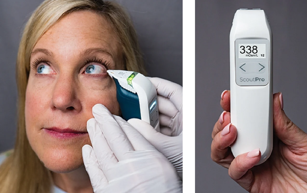
Measures tear osmolarity — the saltiness of your tears — from a sample smaller than a pinhead. It is the closest thing dry eye has to a laboratory number, and it rises as the tear film becomes unstable.
What this means for you: dry eye symptoms are a poor guide to dry eye severity. Some people with badly compromised tear films feel very little, and some people with mild disease are extremely uncomfortable. An objective number tells us where you actually sit, and repeating it tells us whether treatment is working rather than whether you happen to be having a good week. Osmolarity is measured in our Dry Eye Evaluation — a paid, in-depth assessment, with the fee confirmed when you book.
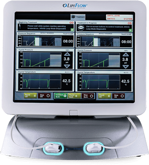
Treats blocked meibomian glands — the oil glands in the eyelids whose secretions stop your tears evaporating. LipiFlow warms the inner surface of the lid, where the glands actually sit, while applying gentle pulsed pressure from the outside to clear the blockage. Warming from the inner surface is the distinction: a hot flannel warms the skin, not the gland.
What this means for you: meibomian gland dysfunction is the most common cause of dry eye, and blocked glands that stay blocked eventually stop working altogether. Treatment is aimed at clearing them while there is still gland to save, which is why we image the glands before deciding. Suitability is decided at a Dry Eye Evaluation first. Read more about our dry eye clinic.
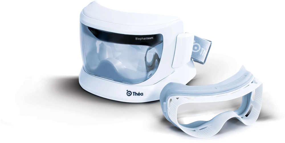
Controlled-humidity warming goggles that hold the eyelids at a steady therapeutic temperature and humidity for a set period, softening hardened oil in the meibomian glands so it can be expressed. It does what a warm flannel is meant to do, but at a temperature that is actually maintained rather than lost within a minute.
What this means for you: it is comfortable, drug-free, and often used alongside IPL and gland expression as part of a course rather than on its own. Which combination you need is decided by measurement, not by symptoms. Start with a Dry Eye Evaluation.
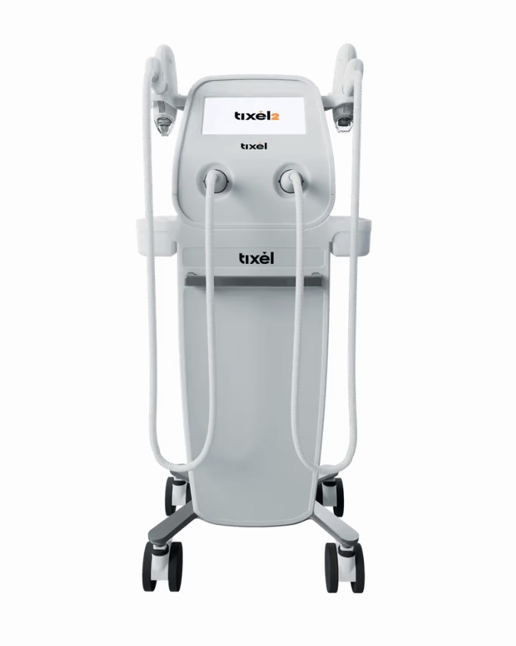
A thermomechanical device that delivers controlled heat to the eyelid skin through a matrix of fine pins, without a laser and without breaking the surface. It is used around the lids to improve gland function and to treat the lid skin itself.
What this means for you: it is another option in a course of dry eye treatment rather than a stand-alone answer, and whether it suits you depends on what your assessment shows. Suitability is decided at a Dry Eye Evaluation.
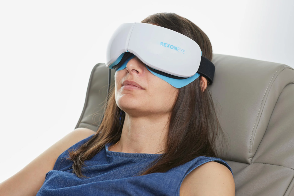
Quantum molecular resonance therapy, delivered through a mask worn over closed eyes. It applies a low-level electrical field to the whole ocular surface and the lids, aimed at the tissue itself rather than at the oil glands alone — which is why it is generally reserved for the more severe and treatment-resistant cases, including inflammatory and aqueous-deficient dry eye.
What this means for you: if you have been through drops, warm compresses and gland treatments without lasting relief, this is one of the reasons it is still worth being reassessed rather than accepting that nothing works. A course is decided after a Dry Eye Evaluation. Read more about our dry eye clinic.
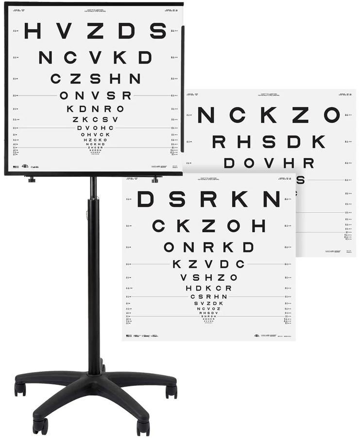
The ETDRS chart is the research standard for measuring visual acuity — the chart used in the clinical trials that establish whether eye treatments work — read under standardised illumination so that the lighting is the same every time.
What this means for you: an ordinary letter chart is fine for prescribing glasses. It is not precise enough to tell whether vision has genuinely changed by a small amount between visits, because the lighting, the chart and the letter spacing all vary. Measuring to research standard means a change we record is a real change, not a difference in the room. Acuity is measured this way as part of your examination — New Patient (45 minutes) or Existing Patient (30 minutes).
A biomicroscope is the instrument an optometrist uses to examine the eye under high magnification with a controlled slit of light — the front surface, the lids and lashes, the lens inside the eye, and, with additional lenses, the retina. Every consulting room at Rose is fitted with a current-generation LED biomicroscope, several with integrated photography, so findings are recorded and compared rather than described from memory.
Alongside them sit digital phoropters, automated lens analysers for reading your existing glasses, and digital acuity charts. None of this is exotic, and that is rather the point: it is the equipment used dozens of times a day, and keeping it current is what makes a routine examination consistent from room to room and from year to year.
What this means for you: the same standard of examination whichever of our optometrists you see, and a photographic record of anything found on the front of your eye so that next year’s appointment starts from evidence rather than recollection. Book a New Patient examination (45 minutes) or an Existing Patient examination (30 minutes).


Frequently asked questions
At Rose, a standard adult eye examination is $129. A new patient appointment takes 45 minutes, and imaging and a full pre-examination workup are included in that price rather than added afterwards. Children and school students are $79, tertiary students $99, and an acute appointment for a sudden problem — a red eye, a foreign body, a sudden change in vision — is $69. Where further testing is needed it is quoted before it is done: visual fields $69, macular OCT $69, full OCT $194, anterior segment OCT $39, corneal topography $59, optical biometry $49, cycloplegic refraction $59. Specialist appointments are priced separately. A keratoconus consultation is $139 for 30 minutes or $169 for 45. An ortho-k consultation is $129. A full dry eye evaluation, which produces a written treatment plan, is $247 — it assumes a current full eye examination, either with us ($129) or a full examination record from another practice within the last six months. Fees elsewhere in New Zealand vary widely, and a lower advertised price usually means a shorter appointment with imaging charged as an extra or not offered at all. It is worth comparing what is included rather than the headline number. Ring us on 07 847 3195 if you want the fee for your situation confirmed before you book.
No practice can honestly claim that, and you should be wary of any that does — "best" depends entirely on what your eyes need. If your eyes are straightforward, several good practices in Hamilton will look after you well, and we would rather say that than pretend otherwise. What separates practices is what they can measure, and what they can treat when things are not straightforward. Rose was built around the complicated end. The Rose K lens for keratoconus was designed in this practice, and lenses of that family are, on the manufacturer's figures, among the most widely prescribed in the world for the irregular cornea. We hold swept-source OCT, corneal tomography, epithelial thickness mapping, two independent optical biometers for monitoring a child's short-sightedness, and objective tear film assessment. For dry eye we hold intense regulated pulsed light, LipiFlow, Blephasteam, Tixel and Rexon-Eye — to the best of our knowledge no other practice in New Zealand has all of these under one roof. A better question than "who is best" is "who is set up for what I have". Ask any practice you are considering how long the appointment is, what they can measure, and whether imaging is included or extra. Those answers will tell you more than a ranking would.
Not usually as a full examination, but there are genuinely free and subsidised pathways worth knowing about. Pre-school children are screened free through the B4 School Check, and there is vision screening in schools. Both are screenings rather than eye examinations — they are designed to catch obvious reduced vision and they miss a great deal, including focusing problems and early short-sightedness. A child who passes a screening can still need glasses. Children under 16 whose family holds a Community Services Card, or who hold a current High Use Health Card, may be eligible for the government spectacle subsidy towards an examination and glasses. MSD criteria apply, and we accept MSD and WINZ payment cards. Southern Cross members can use their optical benefits with us, and we offer Afterpay, Genoapay, QCard and GoCardless. We also run two free 15-minute chats. The myopia chat is for parents of a short-sighted child who want the options explained and want help deciding what is actually right for their family, with our experience behind that decision rather than a brochure. The ortho-k chat is for anyone considering overnight lenses who wants to understand what is involved before committing. Neither is an eye examination, but both are a proper conversation with someone who does this work. If cost is the reason you have been putting off having your eyes checked, ring us on 07 847 3195 and say so. We would rather find a way than have you not come.
While payment is generally required on the day of appointment, we do offer a number of payment options including Afterpay, Genoapay, QCard, and GoCardless.
No. We are closed on public holidays and long weekends as we believe our staff should have the chance to enjoy this time with friends and family.
If your eyes are straightforward, almost any good optometrist can look after you well, and we would rather say that than pretend otherwise. Come to us when something is not straightforward: a prescription that will not settle, a cornea that has changed shape, a child whose short-sightedness is moving faster than it should, an eye surface that has stopped being comfortable, or a diagnosis you have been given but do not understand. Those problems are decided by measurement, and measuring them properly needs instruments most practices do not have.
Yes, and you do not need to justify it. A second opinion is not a criticism of whoever saw you first — very often they were right, and what you actually needed was the finding explained and the numbers put in front of you. What we can add is measurement. We can image the layers of your cornea and retina, map the epithelium, quantify how far your cornea sits from normal, measure the length of your eye and check your tear film objectively. That turns “something is not right” into a number, and a number either explains the problem or rules it out. Bring any previous scans, prescriptions and letters. Comparison against your own past results is often the most valuable thing in the room.
Usually. Complex is what this practice was built around — keratoconus, corneal grafts, irregular corneas after surgery or injury, severe dry eye, high prescriptions, and eyes that have been told they are not fittable with contact lenses. The Rose K lens for keratoconus was designed in this practice, and lenses of that family are, on the manufacturer's figures, among the most widely prescribed in the world for the irregular cornea. If we cannot help, we will tell you at the first appointment and tell you who can, rather than taking you through a course of treatment that was never going to work.
Waiting is the one thing that cannot be undone. Short-sightedness is axial — the eye is physically growing too long — and that length does not come back. What treatment does is slow the rate of growth, so the earlier it starts the more length is saved. We measure axial length directly on two independent biometers and plot it against normal growth curves, which means we can see a child drifting off their expected curve before the prescription changes. There is a free 15-minute myopia chat if you want to understand the options before committing to anything. It is worth being precise about what we are all trying to achieve. Myopia control does not stop a child's eyes changing and no practice should tell a parent it will. What it does, reliably and across many published trials, is slow the rate at which the eye lengthens — and because the risk of retinal detachment, myopic maculopathy and glaucoma in later life rises with every extra millimetre, slowing that growth is how a child is kept out of the range where sight is genuinely at risk. After seventeen years of focusing on this, we are now seeing children on customised plans whose axial length is not progressing at all over a monitoring period, and in some cases measurements that come back slightly shorter than the ones before them. That is our own clinical observation in our own patients rather than a trial result, and it has to be read carefully, because the choroid thickens when treatment is working and part of an apparent reduction can be that thickening rather than the eye itself shortening. That is precisely why we measure both. We are not yet able to tell a parent that we can stop myopia in its tracks, and we will not say it until we can.
Two things: we measure it before treating it, and we hold every current treatment technology in one building. Most dry eye is treated on the basis of symptoms, which is why drops so often fail. A tear film can be failing because the oil layer is blocked, because the water layer is short, because the surface is inflamed, or because the lids are the problem, and those need different treatments. We assess the tear film objectively with TearCheck — tear film stability, the oil layer, lid and gland imaging, and an inflammation measure — and measure tear osmolarity, the saltiness of the tears, which is the closest thing to a laboratory number for dry eye. Then we treat the cause. Rose holds intense regulated pulsed light for inflammation and gland function, LipiFlow thermal pulsation to clear blocked meibomian glands from the inner lid surface, Blephasteam controlled-humidity warming, Tixel thermomechanical treatment for the lids, and Rexon-Eye quantum molecular resonance for the more severe and treatment-resistant cases. At the time of writing, and to the best of our knowledge, no other optometrist or ophthalmologist in New Zealand has all of these available under one roof. We say that as a statement of fact rather than a boast, and we would be glad for it to stop being true. Dry eye is one of the most common and most under-treated conditions we see, and there are far more people living with it than one practice can look after. Any practice wanting to build this kind of service has our encouragement and our help — please contact us for co-management, and send us the cases you are not yet set up for. Why holding all of them matters is simple. A practice with one dry eye machine will offer you that machine. Holding all of them means the assessment decides the treatment, rather than the equipment deciding it — and when the first approach does not work, the next one is in the next room rather than in another city.
Yes — from around six weeks of age. Babies and toddlers are tested with Cardiff acuity cards and retinoscopy, neither of which needs the child to read a letter, name a picture or say a word. Most of the findings that matter in a young child cause no symptoms and are invisible to a parent, which is exactly why they need looking for.
In NZ an optometrist can calculate the prescription for glasses, fit contact lenses, diagnose and treat eye conditions and infections with medication. They can also manage ocular injuries including removing foreign objects. If surgery or specialist treatment is required they can refer to an ophthalmologist (eye surgeon), hospital, or other health care specialist. NZ-trained optometrists must complete a 5-year degree at the University of Auckland before becoming registered.
A dispensing optician has formal training in optical dispensing. They are qualified to help a client choose frames, read a prescription, discuss lens options, take measurements, order and fit glasses.
An ophthalmologist is a medical doctor who has specialised in the treatment of serious eye disease and ocular surgery such as cataract removal. They do not typically prescribe glasses or contact lenses.
Not always, and the difference is usually time and instruments rather than the optometrist. A low advertised fee generally buys a shorter appointment aimed at producing a spectacle prescription. That is a legitimate service and for many people it is enough. What it often does not include is retinal imaging, OCT scanning of the optic nerve and macula, measurement of eye length, or objective assessment of the tear film — the tests that find glaucoma before you notice it, detect macular change early, monitor a child's short-sightedness properly, or explain why your eyes feel gritty. At Rose a new patient appointment is 45 minutes, and imaging and the full pre-examination workup sit inside the $129 rather than on top of it. We are not the cheapest and do not try to be. Whichever practice you choose, three questions are worth asking: how long is the appointment, is imaging included or charged separately, and what happens to the images afterwards. A scan that is taken and kept becomes the baseline every future scan is compared against, and that comparison is frequently worth more than any single result.
Start with an optometrist in almost every case. An optometrist is your first point of contact for eye health and vision — examining the eyes, prescribing glasses and contact lenses, diagnosing eye disease, and in New Zealand treating many conditions with prescription medicines. An ophthalmologist is a medical doctor and eye surgeon who treats serious disease and performs surgery such as cataract removal, and who does not usually prescribe glasses or contact lenses. Neither is better than the other; they do different jobs. Most eye problems are diagnosed and managed by an optometrist, and where surgery or specialist medical treatment is needed we refer you promptly and stay involved in your care. The exception is anything sudden and severe: sudden loss of vision, a curtain across your vision, severe eye pain with a red eye, or a shower of new floaters and flashes. Phone us on 07 847 3195 rather than booking online — a person, not a calendar, decides how quickly you are seen — or go to hospital.
We are at 38 Lake Road in Frankton, with parking on site, a few minutes from the city centre and straightforward to reach from the western side of Hamilton. Our patients come from right across the city — Frankton, Dinsdale, Nawton, Te Rapa, Glenview, Hamilton East, Hillcrest, Chartwell, Rototuna and the central city — and from across the wider Waikato, including Cambridge, Te Awamutu, Morrinsville, Matamata, Huntly, Ngaruawahia, Otorohanga and Raglan. Because much of what we do is specialist work, a good number travel considerably further. We regularly see patients from Auckland, Tauranga, Rotorua, Taupo, Taranaki and the lower North Island for keratoconus, scleral and custom contact lens fitting, and complex dry eye — and we see patients from overseas who combine treatment with a trip. If you are travelling a distance, tell us when you book and we will group your appointments so the journey is worth making.
Forty-five minutes for a new patient, and the appointment is built so the measuring is done before you sit down with the optometrist. A full pre-examination workup and imaging are included in the fee rather than charged on top: assessment of your vision and prescription, examination of the front and back of the eye, eye pressure, and retinal imaging that becomes your permanent baseline. You will leave knowing what we found, what it means, and what — if anything — needs doing. If the honest answer is that nothing needs doing, that is what you will hear. If something needs watching or treating, you get a clear plan and a review date. Where a specific problem needs a specific test — visual fields, OCT, corneal topography, eye length measurement — those are quoted to you before they are done, never added afterwards without your agreement. Existing patients returning for a routine examination are booked for 30 minutes. Please arrive ten minutes early, and bring your current glasses or contact lenses, any recent prescription or referral letter, and a note of the medications you take, because some affect the eyes.
Yes. We keep acute appointments for sudden problems — a red or painful eye, something in the eye, a sudden change in vision, new flashes or floaters — and we see you the same day wherever we can. An acute appointment is $69. Phone first, on 07 847 3195, rather than booking online. A two-minute call establishes whether you need us or the hospital, and a person, not a calendar, decides how quickly you are seen. Any Hamilton optometrist can be phoned for a same-day problem. If we cannot fit you in, we will tell you straight away so you can try the next nearest practice rather than wait. Some things should not wait for an appointment at all. For a chemical splash, rinse the eye with clean water straight away and keep rinsing, then go to the hospital emergency department. For sudden loss of vision, a curtain or shadow across your vision, a penetrating injury, or severe eye pain with a red eye, phone us immediately during opening hours; outside our hours, go to the Waikato Hospital emergency department or call Healthline on 0800 611 116 for advice. You do not need to be an existing patient, and you do not need a referral.
See things the Rose way
Make an appointment at Hamilton’s leading eye care specialists.

