Sudden Vision Loss with Keratoconus? It Could Be Corneal Hydrops
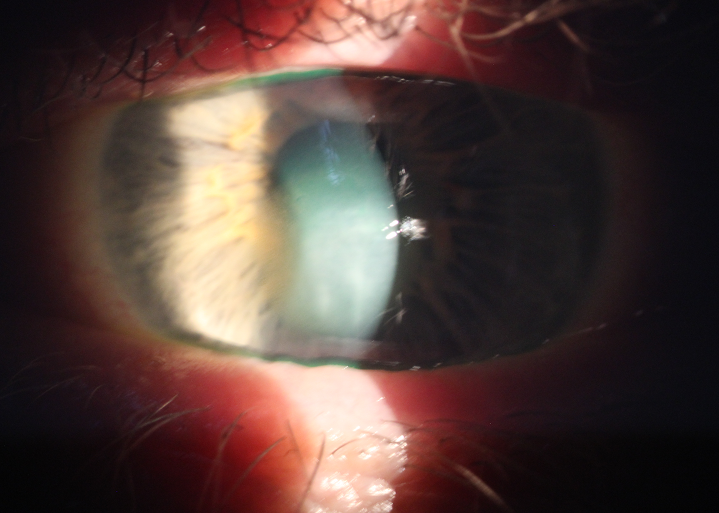
What is Acute Corneal Hydrops?
Acute corneal hydrops is an uncommon complication of keratoconus. As keratoconus progresses, the cornea becomes thinner and weaker. Occasionally, the inner supporting layer of the cornea (Descemet's membrane) develops a tear, allowing fluid from inside the eye to enter the cornea. The result is sudden corneal swelling causing rapid vision loss, glare, discomfort and difficulty wearing contact lenses.
‑

Symptoms That Should Never Be Ignored
- Sudden blurred vision
- A white or cloudy patch on the cornea
- Pain or discomfort
- Increased light sensitivity
- Excessive tearing
- Difficulty wearing contact lenses
- Vision changing dramatically over hours or days
Why Early Assessment Matters
Not every cloudy cornea is hydrops. Corneal infections and other conditions can appear similar but require different treatment. An urgent examination allows confirmation of the diagnosis, exclusion of infection, advanced imaging, monitoring of healing and timely referral when appropriate.
How We Assess Keratoconus
We use slit lamp biomicroscopy, corneal topography, corneal tomography, anterior segment OCT and pachymetry to diagnose and monitor keratoconus and suspected hydrops.
Recovery
Most cases gradually improve over weeks to months. During recovery we monitor corneal clarity, thickness, healing and vision, and many patients can later be successfully fitted with Rose K, scleral, EyeSpace Ectasia or other specialty contact lenses. Keratoconus itself can continue to progress even years after diagnosis — including past age 30 in some people (Gokul et al., British Journal of Ophthalmology, 2017) — which is one more reason we keep monitoring corneal shape at regular reviews long after the initial diagnosis.
Why Choose Rose Optometry
Rose Optometry has cared for patients with keratoconus for decades. Founded by Paul Rose, developer of the internationally recognised Rose K lens, our team continues to contribute to research and innovation through the New Zealand Eye Research Centre (NZERC).
If you have keratoconus and develop sudden blurred vision, pain, light sensitivity or a white patch on your eye, contact Rose Optometry immediately for an urgent assessment. Early diagnosis can make a significant difference to your comfort and long-term vision. Book online.
References
Gomes JAP, Tan D, Rapuano CJ, et al. Global Consensus on Keratoconus and Ectatic Diseases. Cornea. 2015;34(4):359-369. PMID: 25738235.
American Academy of Ophthalmology. Preferred Practice Pattern: Corneal Ectasia.
Maharana PK, Sharma N, Vajpayee RB. Acute Corneal Hydrops in Keratoconus. Indian Journal of Ophthalmology. 2013;61(8):461-464. PMID: 23925338.
Tuft SJ, Gregory WM, Buckley RJ. Acute Corneal Hydrops in Keratoconus. Ophthalmology. 1994;101(10):1738-1744. PMID: 7936572.
Gokul A, Patel DV, Watters GA, McGhee CNJ. The Natural History of Corneal Topographic Progression of Keratoconus After Age 30 Years in Non-Contact Lens Wearers. British Journal of Ophthalmology. 2017;101(6):839-844. PMID: 27729309.
This article is general information, not a substitute for a personalised assessment. If you have keratoconus and notice sudden vision loss, pain or a white patch on your cornea, seek urgent assessment rather than waiting.




See things the Rose way
Make an appointment at Hamilton’s leading eye care specialists.

