OCT Angiography in Hamilton: Seeing the Eye's Blood Vessels Without Dye
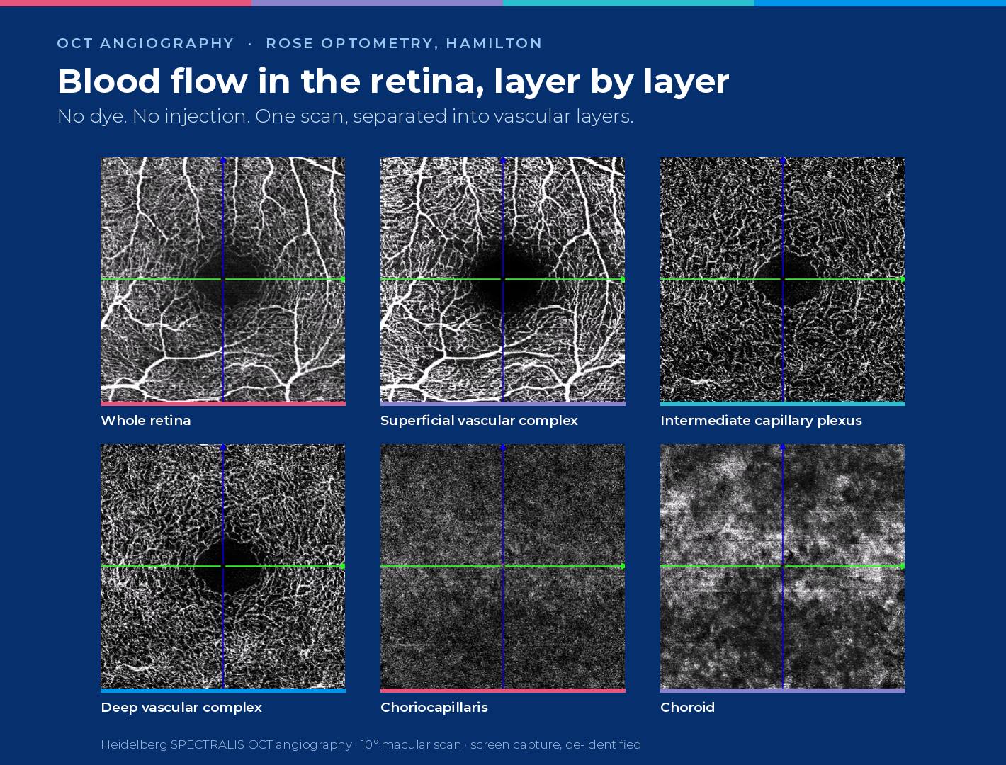
Short answer: OCT angiography (OCTA) is a scan that maps the blood vessels inside the retina and the layer beneath it, the choroid, without any dye or injection. It takes seconds per eye. Rose Optometry in Hamilton uses the Heidelberg SPECTRALIS OCT angiography module to detect and monitor changes in blood flow linked to macular degeneration, diabetic retinopathy, retinal vein occlusion, glaucoma and myopia.
What is OCT angiography?
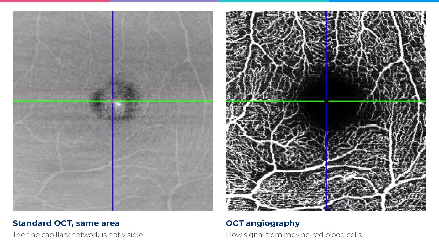
A standard OCT scan builds a cross-section of the retina from reflected light, a little like ultrasound but with light instead of sound. It shows structure beautifully — thickness, layers, fluid — but it cannot tell a blood vessel from the tissue around it.
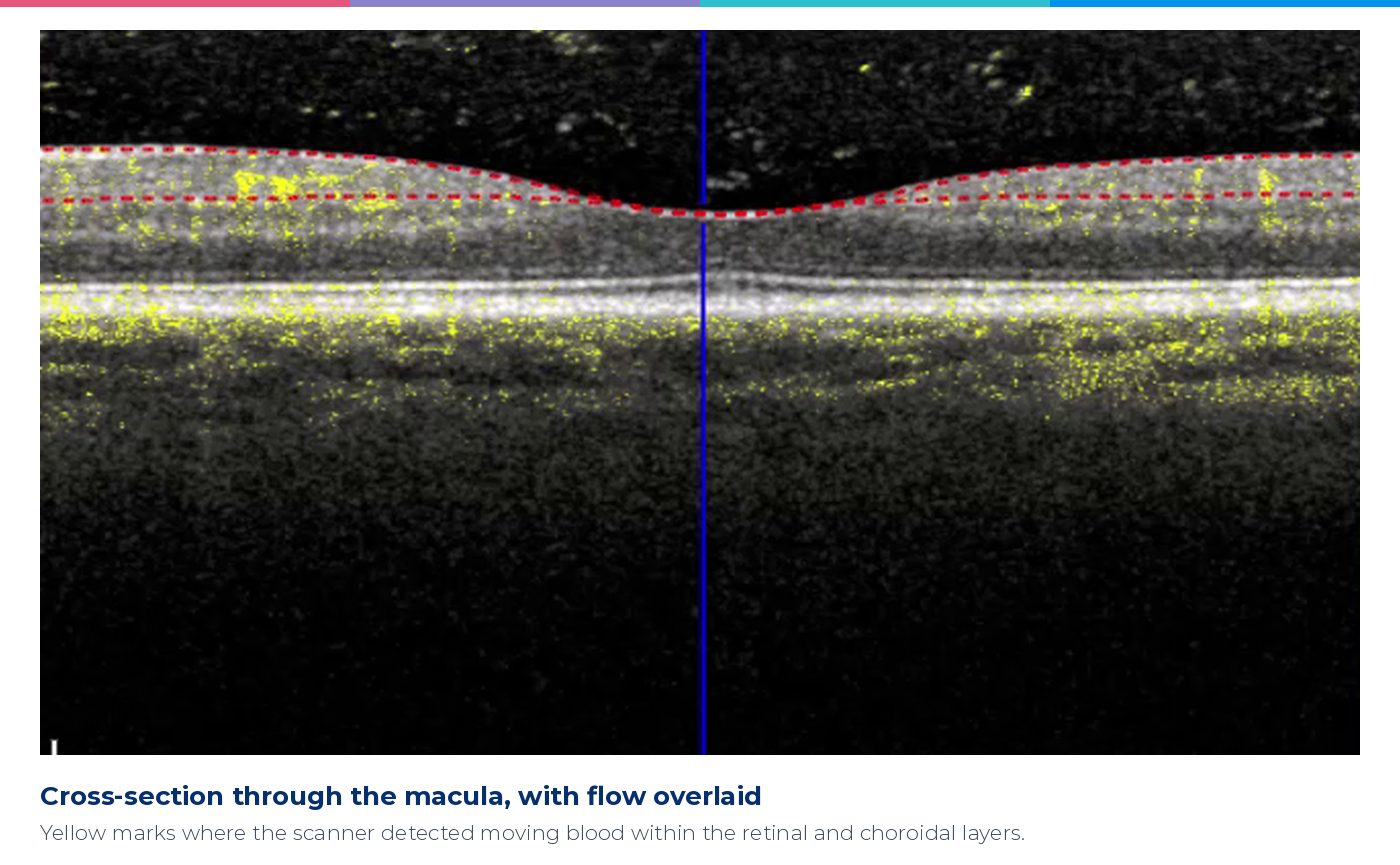
OCT angiography adds movement. The scanner takes several images of exactly the same spot in rapid succession. Anything that stays still looks the same each time; red blood cells flowing through a vessel do not. The software picks out that change and turns it into a map of perfused vessels. Heidelberg's system uses at least four repeated scans per location and up to seven for finer contrast, and its high-resolution scan samples the retina at about 5.7 µm per pixel across and 3.9 µm in depth — fine enough to show capillaries around 8 µm wide.
Because OCT knows depth, the result can be separated into layers. On our platform that means the nerve fibre layer vessels, the superficial vascular plexus, the intermediate and deep capillary plexuses, the avascular outer retina, the choriocapillaris and the choroid — each viewable on its own.
How is it different from a dye angiogram?
The traditional way to see retinal circulation is fluorescein angiography: a dye is injected into a vein in the arm and photographed as it passes through the eye. It remains important in hospital eye care, and it shows something OCT angiography cannot — dye leaking out of damaged vessels.
But it involves a needle, takes time, and carries a small risk of reaction, so it is not something you repeat at every visit. OCT angiography needs no injection at all, which means it can be repeated whenever it is useful. That changes what is practical: instead of a single snapshot when something has already gone wrong, we can build a record over time and compare like with like.
Why this matters in Hamilton
As far as we are aware, Rose Optometry is the only optometry practice in the North Island with OCT angiography. We chose the Heidelberg SPECTRALIS platform deliberately: it is the same Heidelberg imaging platform used by Waikato Hospital's eye department and by Hamilton's private ophthalmologists.
That has practical value for patients. When we refer you, the images we send are from the same family of instrument your ophthalmologist uses, so they can be read and compared directly rather than treated as a separate, unfamiliar dataset. And when you come back to us for monitoring, we are measuring on the same terms as the specialist who may see you next.
Conditions we use OCT angiography to monitor
Age-related macular degeneration (AMD). The event that matters most in AMD is the growth of abnormal new vessels under the retina, called macular neovascularisation, which can leak and cause rapid loss of central vision. OCT angiography can show these vessels directly — sometimes before they leak. In a study of 160 eyes with dry AMD whose other eye already had wet AMD, OCT angiography found silent, non-leaking new vessels in 14.4%. Over the following year, 21.1% of those eyes went on to leak, compared with 3.6% of eyes without them — roughly a 15-fold higher risk (de Oliveira Dias et al, Ophthalmology 2018, using a different OCTA device). Finding them does not mean treatment starts; it means closer follow-up and faster referral if anything changes.
Diabetic retinopathy. Diabetes damages the smallest vessels first. OCT angiography shows capillary dropout and enlargement of the vessel-free zone at the centre of the macula, often before the classic signs appear on a photograph. In a prospective study of 205 eyes followed for about two years, a larger vessel-free zone and lower vessel density in the deep capillary plexus predicted which eyes would progress, and improved risk prediction beyond blood sugar, diabetes duration and blood pressure alone (Sun et al, Ophthalmology 2019).
Retinal vein occlusion. After a vein blockage, OCT angiography maps the areas of retina that have lost their capillary supply, which helps judge the risk of complications and track recovery.
Glaucoma. Glaucoma is usually monitored with visual fields and OCT nerve-fibre measurements. In advanced glaucoma the nerve fibre layer can thin to a floor where further loss is no longer measurable. Vessel density around the fovea keeps falling well beyond that point: one study of 509 eyes found no detectable measurement floor for it, making it a promising way to follow late-stage disease when the usual measures run out (Moghimi et al, Ophthalmology 2019). It complements fields and OCT; it does not replace them.
Myopia and red light therapy. The choroid, the vascular layer under the retina, is central to how myopia develops, and it is the tissue low-level red light therapy is thought to act on. We monitor every red light therapy patient with choroidal thickness measurements, OCT angiography and choroidal vascularity index, on top of OCT and retinal photography. In people with high myopia, OCT angiography can also help identify myopic neovascularisation, a cause of sudden central blur.
Other macular conditions. OCT angiography is also useful in central serous chorioretinopathy, macular telangiectasia and other conditions where the question is whether blood flow is normal, reduced or abnormal.
What OCT angiography cannot do
It is a powerful scan, not a complete answer. It does not show leakage the way dye angiography does. It covers a smaller area of the retina than a wide-field photograph. It needs steady fixation, and larger surface vessels can cast ‘shadow’ artefacts onto deeper layers, which the software reduces but cannot always remove. Results always have to be read alongside your examination, your other scans and your history, and some findings still need a hospital dye angiogram.
What to expect
You sit at the instrument and look at a target while the scan runs — a few seconds for each eye, with nothing touching the eye and no injection. Depending on your pupils and what we are looking for, we may use dilating drops, which blur near vision for a few hours. The images are stored so the next scan can be compared with this one point for point.
Common questions
Is OCT angiography safe?
Yes. It uses the same low-power near-infrared light as a standard OCT scan, with no dye, no injection and no X-rays or other ionising radiation.
Does OCT angiography replace fluorescein angiography?
Not entirely. It avoids an injection and can be repeated freely, but it cannot show leakage. Your ophthalmologist may still request a dye angiogram in some situations.
Who should have OCT angiography?
It is most useful for people with, or at risk of, macular degeneration, diabetic eye disease, vein occlusion, glaucoma or high myopia, and for children on red light therapy. Your optometrist will recommend it when the result would change how your eyes are monitored.
Can my OCT angiography images be shared with my eye specialist?
Yes. Because we use the Heidelberg platform also used at Waikato Hospital and by Hamilton's private ophthalmologists, the images can accompany a referral and be compared directly with the specialist's own scans.
Our optometrists
OCT angiography is part of the clinical practice of all of our therapeutically qualified optometrists: partners Jagrut Lallu and Jacqueline Rowe, and optometrists Emilie Lawson (Accredited Glaucoma Prescriber), Jason Shen, Anjali Hira, Stella Wong and Jessica Wood.
Book a comprehensive eye examination with our team: new patients book here or existing patients book here. Already under our care and due for repeat imaging? Book your OCT appointment here. If you notice sudden distortion, a new grey patch or a rapid drop in central vision, phone us the same day on 07 847 3195.
This article is general information, not a substitute for individual advice. Images are de-identified screen captures from our Heidelberg SPECTRALIS. Sources: Heidelberg Engineering, SPECTRALIS OCTA — Principles and Clinical Applications; de Oliveira Dias JR et al, Natural history of subclinical neovascularization in nonexudative AMD using swept-source OCT angiography, Ophthalmology 2018;125(2):255–266, doi:10.1016/j.ophtha.2017.08.030; Sun Z et al, OCT angiography metrics predict progression of diabetic retinopathy and development of diabetic macular edema, Ophthalmology 2019;126(12):1675–1684, doi:10.1016/j.ophtha.2019.06.016; Moghimi S et al, Measurement floors and dynamic ranges of OCT and OCT angiography in glaucoma, Ophthalmology 2019;126(7):980–988, doi:10.1016/j.ophtha.2019.03.003; Rao HL et al, Optical coherence tomography angiography in glaucoma, J Glaucoma 2020;29(4):312–321, doi:10.1097/IJG.0000000000001463.
Recent articles


See things the Rose way
Make an appointment at Hamilton’s leading eye care specialists.

